Making teeth transparent

Dental imaging is advancing all the time, but despite its growth, there’s still one distinct limitation: The final image often has to be cut into sections to be viewed. In essence, it’s 2D.
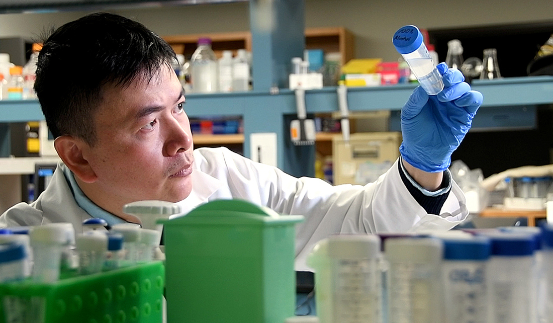
Dr. Hu Zhao, a restorative sciences faculty member at Texas A&M College of Dentistry, considered one workaround. What if the biopsy or organ sample could be rendered completely transparent? While the concept has all the makings for a sci-fi flick, this technology is not new. Zhao, a practicing dentist and researcher, simply adapted a tissue-clearing technique currently used in neuroscience.
“Now we can bring the entire head under the microscope without cutting the section: the nasal cavity, tooth germ and brain,” Zhao says of his patented method, dubbed PEGASOS — polyethylene glycol-associated solvent system — which utilizes images generated from a confocal microscope. “We are the first lab in the world to bring this technique to the dental field. Previously there was no way to make bone transparent.”
Zhao and his research team’s work recently was bolstered with a $400,000 grant from the National Institutes of Health – National Institute of Dental and Craniofacial Research as well as publication in the May 2018 issue of Cell Research.
It’s likely because the shift from opaque to transparent opens up a whole new world.
“We can literally outline every neuron to see how they travel,” says Zhao. “There is no way to get this information from sections. We show the nerves within the bone; everybody knows that there are nerve fibers within the bone marrow, but this is the first time that we can see them. We just make the bone transparent, and image the whole sample as one unit.”
While his lab is just halfway through the two-year grant already, potential applications in dental specialties are becoming clear. Take, for instance, dental implants. With the tissue-clearing technique, samples from mouse models reveal the interface between the implant and the bone down to every last blood vessel.
“You can see the bone directly contacting the implant,” Zhao explains. “There is no other technique that can visualize this.”
Word of the findings has spread not only to collaborators at other institutions, including UT Southwestern, Caltech and Johns Hopkins, but also to other departments closer to home.
Dr. Elisabeth Barnhart, a resident in the graduate orthodontic program, utilized the technology for a portion of her master’s thesis. The goal: View the penetration of an orthodontic sealant into etched enamel. The means: Use the tissue-clearing technique to aid in removing the mineralized tissue in order to better view the sealant.
“We viewed the sample to see if the method was causing dissolution of the sealant from the tooth enamel prior to the entire clearing protocol being completed,” says Barnhart. “The sealant was in fact still present in the samples, which shows promise for future studies using the technique.”
There is potential in diagnostics and pathology, as well.
Much of Dr. Lisa Cheng’s research is devoted to developing a test using salivary biomarkers indicative of oral cancer and other health conditions.
“One future research direction for Dr. Zhao’s novel technique,” says Cheng, director of oral pathology, “is to use it for investigating cancer behavior, specifically how cancer cells spread in the local tissue, spread through nerves, or invade the blood vessel to metastasize.”
With the patent for the PEGASOS method submitted and trademark protections in place, Zhao’s team looks toward creating the next generation tissue-clearing technique while identifying more clinical applications for the technology.
“We are so excited about this,” says Zhao.