This omission is no mistake

There’s just something special about FAM20B. Or at least that’s what early findings indicate. A member of the protein family FAM20, this particular gene is necessary for cartilage development. So it’s interesting to note what happens to teeth when FAM20B is selectively nullified, or removed, from mouse models: The enamel appears to reach a higher state of mineralization. Microscopic analysis reveals the details.
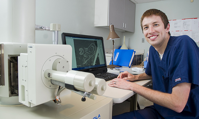
“If it was less mineralized there would be more carbon, and if enamel was more mineralized there would be more calcium, oxygen and phosphorous and less carbon,” explains Derrick Crawford, second-year dental student, who worked last summer with faculty mentor Dr. Xiaofang Wang, assistant professor in biomedical sciences.
What’s more, this faculty-student research team made an eye-opening observation: the growth of additional teeth.
Crawford shared these findings in a fall 2015 research competition at Texas A&M University Baylor College of Dentistry for the American Association for Dental Research Student Research Award. As the first-place winner, he received a $500 travel award and free registration from AADR to attend its annual meeting, which is March 16-19 in Los Angeles. Next week he’ll share these results again, this time with peers and the national dental research community.
While Crawford’s work with Wang lasted several months, there is still much testing to be done. First and foremost, Wang must analyze the enamel in the models to determine quality; namely, its structure, hardness and resistance to decay.
“It will be very exciting if there is a way to improve the quality of enamel in the lab, and the supernumerary (extra) teeth phenotype of Fam20B knockout mice is even more provoking for us, because it clearly shows potential for tooth regeneration; however, this will need tremendous work before applying to humans,” says Wang.
As an incoming third-year dental student, patient care in the clinic starting this summer will take Crawford from the research bench to the dental operatory, but the relevance of the experience to his dental education has not lost its impact.
“The scanning electron microscope images that I made were of tooth enamel and dentin,” says Crawford. “We learn about the physical makeup of these crystals and how they form the enamel rods, and how they fit together, but to be able to look at that firsthand and zoom in and see the elemental makeup — not just a picture in a PowerPoint presentation but something I’ve taken and polished and put into a microscope — brings a whole new application of mineralization and demineralization.”