Unlocking mysteries of the past with the materials of today
Prehuman fossils, diet and the surprising correlation with craniofacial development
There’s been a lively debate floating among biological anthropology circles in recent years, and at the center of it is Australopithecus sediba, a prehuman species that lived two million years ago in southern Africa. While researchers agree on the probability that Australopithecus sediba is a close relative of early humans, there is one important unresolved matter: this species’ diet.
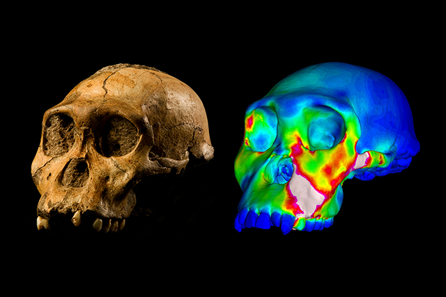
After the 2008 discovery of an Australopithecus sediba fossil skull in Malapa, a cave near Johannesburg, South Africa, the discussion generated international attention in 2012 when research was published suggesting this species lived on a diet rich in tree bark, plant products and fruit.
Findings published Feb. 8 in Nature Communications render that theory quite improbable. An international team including researchers from Texas A&M University Baylor College of Dentistry shows that Australopithecus sediba just didn’t have the jaw and tooth structure necessary to eat a diet of hard foods on a consistent basis.
What’s interesting to note is that while Australopithecus sediba demonstrated limitations in biting, as we do, other prehuman species weren’t as limited in this regard. This points to the possibility that some australopith populations evolved to maximize their bite, while others moved in the opposite direction.
“This has been a hot topic lately, how diet and feeding in these animals affects the skull morphology, and how what kinds of foods they ate drove craniofacial evolution,” says Dr. Paul Dechow, Regents Professor and chair of TAMBCD’s Department of Biomedical Sciences, one of the principal investigators on the project.
Dechow worked with Dr. Qian Wang, associate professor, Leslie Pryor, graduate student in biomedical sciences, and an international team of scientists. Together they obtained information on the bone structure and function of modern primate skulls using ultrasonic techniques and finite element modeling, biomechanical methods akin to the ones utilized by engineers to test whether planes and cars are strong enough to withstand use without breaking. They applied loads to primate skulls and then measured the strains on the skulls to see if they were similar to those predicted by the model. The data was used to validate finite element models of the Australopithecus sediba skull.
TAMBCD researchers are uniquely positioned to contribute to research of this nature because Dechow’s lab produces 3-D material properties of craniofacial skeletons, the only lab of its kind in the world to do this. Dechow and Wang’s studies of bone material properties in monkeys, humans and great apes indicate that elastic properties of craniofacial bone can vary according to anatomical regions, yet closely related species have similar patterns of variation, which provides fundamental basis of using chimpanzee data for building up realistic models of human ancestors with confidence.
So just how does understanding the eating habits and masticatory function of prehumans millions of years removed benefit the dentists of today?
“The whole study of human evolution and all the craniofacial changes that took place in human evolution are important for understanding the current structure and mechanics of our dentition and chewing apparatus,” Dechow says. “It is particularly important for a number of problems associated with the modern human dentition, modern human malocclusion being a very important one.”
It also identifies a shared concern: capability of the temporomandibular joint, or TMJ.
“The TMJ was the weakest link from early on,” says Wang, one of several principal investigators on the international research team. Perhaps the most striking part of their findings: “Australopithecus sediba seemed to have stronger bone, yet like us, they could not consume very hard food.”
The implications for modern dental study and practice are apparent.
“There is a delicate harmony in the recruitment of muscle forces, positioning of biting points and the function of the temporomandibular joint,” says Wang. “Too much force recruited or positioning the jaw in an abnormal way may put undue stress on the TMJ, compromising the joint.”
Collaboration for the research dates back more than a decade, preceding even the 2008 discovery of Australopithecus sediba. The partnership spans continents — and the state, as Texas A&M professor and biological anthropology program coordinator Dr. Darryl de Ruiter has an active role in the project: He is one of the researchers who discovered and described the Australopithecus sediba skull.
“I worked very closely with Drs. Dechow, Wang and Pryor on the development of this paper, though the expertise in biomechanics lies with them and not me,” de Ruiter says. “Together our team generated the necessary models, and tested them to assess what kind of bite forces Australopithecus sediba would have been capable of generating, and how this relates to its diet.
“For me it was quite an enlightening process, watching how some of the world’s leading biomechanics specialists debate the interpretations of their research results. Being able to conduct interdisciplinary, collaborative research is at the very heart of how we organize Malapa research. No single author could have produced all of the necessary work that went into this paper, therefore were it not for this collaboration, we would not know of the mechanics of chewing in Australopithecus sediba.”