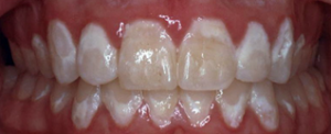
White spot lesions

How do you get orthodontists to research — let alone talk about — this taboo topic? Appeal to their sense of loyalty and form an alumni practice-based research network, of course.
Something started to shift in 1982. That was the year Dr. Phillip Campbell started bonding brackets to his patients’ teeth instead of individually wrapping bands around each tooth.
“Within six months I started to see white spots form, and I was thinking, ‘Whoa, what’s happening?’ From that point forward, it’s been a problem.”
It led Campbell, then practicing in Huntsville, Texas, to do a little office-based research. He reviewed 100 cases of patients whose orthodontic work had been completed with bands and compared them to 100 cases of patients whom he had treated with bonded brackets.
Of the patients treated with bands, only three had small white spots.
“There was just a little line where the cement had washed out under the band, and the tooth had decalcified a little bit,” Campbell explains. In contrast, 21 of the patients treated with bonded brackets had white spot lesions — and the white spots, well, they went all the way to the gum line.
“I started asking myself, ‘What are we doing differently?’” Campbell says. Could it have been the etching with phosphoric acid prior to placing the brackets? Other than that, both methods used cements as dental adhesives.
The answers were a long time in coming, and there was an obvious reason why: He couldn’t get any orthodontic colleagues to talk about it.
“Most of my colleagues would say, ‘I don’t really have a problem with it.’” He wondered, Why are my patients different?
“We all would like white spot lesions to go away. But they’re not going away,” says Campbell, now Robert E. Gaylord Endowed Chair of the orthodontic program at Texas A&M University Baylor College of Dentistry. “We still don’t have all the solutions. We’re working on it. Now most orthodontists will admit that it’s a problem, and it really is disappointing when you work really hard to get the teeth beautiful and straight, and you end up with scars on the teeth.”
While there are options, including using tooth-colored fillings and, more recently, resin infiltration to disguise the telltale milky white spots — essentially cavities on the smooth surfaces of teeth — it’s better to prevent the condition in the first place.

“It’s kind of like you unveil the case, and you see a white spot lesion, and your blood pressure just drops,” says Campbell. “It’s easy for people to say, ‘Well, it’s the patients’ fault, because they didn’t brush properly.’ Well, that’s true, but we’re the treating doctors, and our charge is to do no harm.”
So in 2012, then-orthodontic resident Dr. Matthew Brown made the topic the focus of his master’s thesis.
With help from Campbell and Dr. Peter Buschang, Regents Professor in orthodontics, they solicited alumni to ask a favor: Would they be willing to donate some of their own time and office resources to explore the prevalence of white spot lesions in their own practices?
For alumni like Dr. Ralph Brock ’02, who maintains a practice in Katy, Texas, coupled with a part-time faculty role as assistant clinical professor in orthodontics, the decision was easy.
“Although white spot lesions are not an easy topic to discuss, the orthodontic/dental community can better adapt to the unexpected with the knowledge gained from research of this nature,” says Brock. When it comes down to it, he says it was “the request by my orthodontic program, the prevalence of the white spot lesion concern and my desire to contribute to the understanding of white spot lesions” that motivated him to help with the project.
What’s interesting to note is that in the abstract of the article, “A Practice-Based Evaluation of the Prevalence and Predisposing Etiology of White Spot Lesions,” published in the August 2015 issue of The Angle Orthodontist, 90 percent of the alumni were willing to participate solely based on the fact that a fellow alumnus asked them to.
The research team gauged that interest with a survey. First, says Buschang, respondents said they would be willing to participate — but it would depend on several factors.
“If just anyone were to ask them, they probably wouldn’t do it,” he adds.
Follow-up questions and responses went something like this:
How about if someone from the orthodontic/dental community asked you?
Maybe, but … probably not.
What about if another orthodontist asked you?
Maybe.
How about a fellow alum?
Of course.
It wasn’t a small commitment on the part of the 20 alumni who were selected to participate in the research. These orthodontists were required to obtain informed consent from patients whose cases were used in the study, comb through records to get a feel for oral hygiene and compliance, and even have patients complete questionnaires on the subject.
The results: Of the 158 treated cases analyzed, approximately 28 percent of patients were found to develop white spot lesions. These patients developed, on average, 2.4 white spots, which affected 12.7 percent of teeth examined. Risk factors pointed to poor oral hygiene, poor gingival health and long treatment times.
This mirrors a TAMBCD study led by Dr. Katie Julien ’92, ’94, clinic director and assistant professor in orthodontics, which was published in The Angle Orthodontist in 2013.
Approximately 23.4 percent of 885 orthodontic patients treated at TAMBCD developed at least one white spot lesion during the course of their treatment. The risk factors were nearly identical, with no differences between male and female patients: pre-existing white spot lesions, decline in oral hygiene during treatment, poor pretreatment oral hygiene, and treatment time lasting longer than 36 months.
So what is an orthodontist to do?
“For orthodontists as well as dentists, we need to take this problem seriously,” says Julien. “We need to look at our new patients critically and control our urge to immediately put braces on every child who needs them.”
Her recommendations: Educate the patient and parent, wait until oral hygiene is under control before planning orthodontic treatment, and utilize dental sealants, a protective covering typically placed over the chewing surfaces of teeth to prevent cavities. These can be applied to the entire tooth before treatment and multiple times in high-risk cases. Keeping the patient’s time in braces to a minimum may also help.
“Overall, we need to protect our patients’ teeth with sealants, reapply as needed, watch their level of hygiene at each appointment, educate them and watch our treatment efficiency,” says Julien. “We want to end up with healthy, beautiful smiles in 100 percent of cases.”